Thursday, November 13, 2008

Principle Thoughts about Lenses + Filters for UV

Some priciple thoughts, why normal lenses and filters do not work for reflected ultraviolet (UV) photography.

I get quite a few questions, why a certain camera, lens, filter setup does not or not satisfyingly work for UV photography. I will try and show the effects of that optical system using some transmission graphs I recently made.

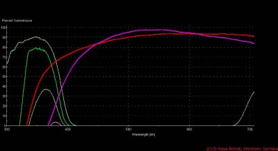
So here some results using one my X-lenses (red line) and a very modern, multicoated lens (purple line), a Baader 2" U-filter (dark green line) and a common UV transmissive filter (very light green line) such as a Schott UG-1, Hoya U-340, B+W 403, Kodak Wratten 18A, Nikon FF etc. sometimes also called "Woods Glass".

Let's look at the filters first. The older, common UV filter has a benefit, quite a high peak UV transmission of about 90% - but it also shows IR transmission from about 670nm onwards. In former times of film based cameras, that was no issue, since these film had no IR sensitivity. Today in the times of digital cameras, and especially the modified ones, their IR sensitivity is quite resp. very high. So that leads to quite some contamination of that supposedly UV image due to "IR leakage". One cure would be to add ("stack") another filter in front which passes UV but blocks or reduces IR. Such a filter would be the Schott blue-green BG-38 or BG-40 filter. But that does not completely cure that problem. The german astronomy company Baader developed because of that fact their Baader-U filter (also called "Venus" Filter, since astronomers use it to make details of the surface of planet Venus visible), with perfectly suppressed IR, as the dark green line nicely proves. This fact made it my "filter workhorse" for reflected UV photography, even if the peak transmission is a bit lower at about 80%.

Now about the lenses. Usually a photographic lens should reproduce visible images, which is the wavelength band of about 400-700nm. This is why lens makers apply a special multicoating (aside from achieving higher light transmission), which usually blocks light outside this visible waveband. The purple line of a recent multicoated lens proves, how succesful this is done today. For UV photography, however, the UV-A band (300 - 400nm) is the interesting one. So using a modern lens and that Baader U-filter leads to that mini UV transmission of about 5% only, which usually causes very long exposure times, grainy images and a lot of frustration for some who wanted to step into that field. Here the wrong lens is the reason for that, not the filter!

One solution would be to get one of the highly specialized $$$$ quartz / fluorite lenses I have reported about here, but this is just for a few who can afford that or have access to one of these lenses - surely a pleasure to use. But the ambitioned amateur or researcher on a tight budget needs a solution too.

This was the reason why I started some reasearch to find lenses which would allow similar image quality, but at affordable cost. So my series of X-lenses was born, the most prominent being the X135 (f=135mm), X50 (f=50mm) and X35 (f=35mm), one of them shown as bright red line which shows quite some more and deeper into the UV reaching transmission, one of them to about 310nm.

The system lens + filter using an X-lens now leads to a much higher UV output to the camera chip (assuming a modern digital SLR camera, which also has to have some UV sensitivity, such as a Nikon D70(s), D40 or modified D200) of about 7 times more, a gain of 3 stops or 3EV in photog speak (this is just about 1 stop less than using the UV Nikkor!). This allows much shorter exposure times, better quality in terms of graininess (noise) and UV reproduction, since also a broader UV waveband is covered.

Just a remark, the high UV transmission is a must for a suitable UV lens, but also good sharpness, high contrast, flare resistance, no hot-spots, etc. are requirements such an X-lens has to meet, and I have not jet mentioned an important point - focus shift. This latter point was solved by calibrating such a lens for UV use, to allow for a sharp visible and UV focus.


[click on image let's you see a larger image]


The explanation is not yet complete, two important parts of the system are not covered here in detail, the camera and the light source(s), which should be used for UV photography. I will elaborate on that at a later time here.

Stay tuned, more will follow on that fascinating subject...

More info on this very interesting field may be found on my site http://www.pbase.com/kds315/uv_photos
Tuesday, November 11, 2008

Teeth and UV I

Today about teeth and ultraviolet photography.

I found in some older books some contributions of Prof. Woods and esp. Ch. Lunnon about photograping teeth using ultraviolet light.

So here some results using my calibrated for UV X35 lens, a Baader 2" U-filter and my newly developed High Power UV flash.

The top image clearly shows, repairs, cracks, plaque etc. whereas the second one using a special filtering technique also shows bacteria build up (plaque) as reddish cast and repairs having being done. The small size of the images here do not really show the amount of detail which gets visible using these techniques.

[click on image let's you see a larger image]


Stay tuned, more will follow on that fascinating subject...

More info on this very interesting field may be found on my site http://www.pbase.com/kds315/uv_photos

Wednesday, October 1, 2008

Lens Comparison: UV Nikkor 105mm, COSI 105mm, B&L 9"

Today I got an old lens, which was made many years ago by famous lens maker Bausch & Lomb, Rochester NY. It is a golden color single coated, heavy and well made f4.5 9" lens which attracted me, since it was made to record images emitted by a P-16 phosphor screen. Nothing special actually, but P-16 radiates at about max. 380nm, so now you know what attracted me most...

I ran a quick test using my "standard test subject", a roof of the neighbours house against the Coastal Optics Micro Apo 105mm and the Nikon UV Nikkor 105mm. The B&L lens results are shown on the right side of the comparison images. The B&L images have been scaled down to match the shorter focal length of the other two lenses. These are 100% center crops. The Baader U-filter (310 - 390nm) was used for the reflected UV shots. [click on image gets you a larger image]

Here the results in visible light for the UV Nikkor (left):


and for the Coastal Optics 105mm (left):


Here the results in reflected UV light using the Baader U-Filter (310 - 390nm) for the UV Nikkor (left):


and for the Coastal Optics 105mm (left):


So now it is up to you to understand my surprise, how amazingly sharp this B&L lens is....

Stay tuned, more will follow on that fascinating subject...

More info on this very interesting field may be found on my site http://www.pbase.com/kds315/uv_photos

Allium (Onion) in UV

So this is about onions in UV light. These have very cute ball-shaped flowers and I wondered how these would look like in UV "light". The petals seem to have quite some strong UV reflectance to attract pollinators.

So have a look, left is the visual image, middle the reflected UV image and right the image with UV remapped into the visual as blue [click on image gets you a larger image]:


And this one in a bit higher resolution:


So I hope you like these as much as I do....

Stay tuned, more will follow on that fascinating subject...

More info on this very interesting field may be found on my site http://www.pbase.com/kds315/uv_photos

Saturday, September 20, 2008

UV Images Exhibit in Weinheim

So this is about my upcoming next UV picture exhibit which will be opening September 20st, 2008 in Weinheim, Germany in cooperation with very creative and esthetic Christian Mayer Floristik. That same weekend "Weinheimer Hebst" ("Autumn in Weinheim") will take place, with all shops open during the whole weekend. (No big deal you think? Not in Germany I tell you...)

So have a look, these images will be on display there for a few weeks [click on image gets you a larger image]:


and this is how it looks now there:


[exhibit ended Nov 7th, 2008]

So I hope you like these as much as I do....

Stay tuned, more will follow on that fascinating subject...

More info on this very interesting field may be found on my site http://www.pbase.com/kds315/uv_photos

Friday, September 5, 2008

UV and Forensic Skin Photography I

This will be the start of some entries about forensic photography. Using different wavelengths, filters, lenses and special lights, I would like to show, what may be documented on human skin using these techniques.

Lets start documenting some bruise and its development over time. Reflected ultraviolet photography is said to be able to detect these bruises even if they have disappeared in the visual. So let's see....

Here now the first image set, top row is bruise after + 4 days, second after + 8 days. Color is useless in this content I thought, so it will be "just" black and white images. I will elaborate on the different filters and wavelengths used later. 
[click on image to get a larger one]


And here how that looks like 44 days (1.5 months) later, it gets clearly visible that the normal, visible light photography hardly picks up anything of that healed bruise, whereas the special setup used (image on the right) still clearly shows that old, healed bruise!


And this is how "invisible" older and already healed scratches may be made visible:


...more will follow on that.


So I hope you like these as much as I do....

Stay tuned, more will follow on that fascinating subject...

More info on this very interesting field may be found on my site http://www.pbase.com/kds315/uv_photos

Monday, September 1, 2008

UV Fluorescent Minerals

Today now very bright images which fluorescent minerals can provide. A very good friend of mine was so kind to open up his mineral collection "vault" for me and I was able to take some shots of his shotwave and longwave fluorecent mineral collections. Since he has the needed ultraviolet lamps already installed in his cabinets, a "normal" camera and lens was sufficient, tripod mounted because of the somewhat longer exposure times.

Here now the images, click on them to get larger ones:


and this now an also fluorescent shrimp:


So I hope you like these as much as I do....

Stay tuned, more will follow on that fascinating subject...

More info on this very interesting field may be found on my site http://www.pbase.com/kds315/uv_photos

Tuesday, August 26, 2008

Literature on UV (and IR) Photography

Now this one won't contain pictures and new findings, but a list of literature which deals with UV photography. I have collected that over the years and I am very greatful for the work of Dres. Williams who have provided also quite a few useful resources which I will happily included also.

So here it is:

Books:
1) Quirke, B.A. "Forged, Anonymous, And Suspect Documents", Routledge & Sons, London, 1930 where in chapter XXII on some 19pp ultraviolet and flourescence photography is used to make forgeries visible
2) Matthews S.K., "Photography in Archeology and Art", Humanities Press, London 1968, chap. 12 some 8pp on UV and IR photography
3) Engel, Ch.E. (Ed.) "Photography for the Scientist", Academic Press, London and NY, 1st Edition, 1968, Chapter 8 by Peter Hansell on Ultraviolett and Fluorescence recording, ca 19pp
4) Morton, R.A. (Ed.) "Photography for the Scientist", Academic Press, London and NY, 2nd Edition, 1984, Chapter 8 by Peter Hansell and Raymond J. Lunnon on Ultraviolett and Fluorescence recording, ca 33pp
5) Arnold, Rolls, Stewart, "Applied Photography", Focal Press, London and New York, 1st Ed. 1971, chap. 8 Ultraviolet Photography, 48pp very detailled about lighting, filters, lenses, developping, detectors, applications
6) Rorimer J.J., "Ultra-Violet Rays and their use in the Examination of Works of Art", Metropolitan Museum of Art, New York, 1931 with tons of pictured examples
7) Pountney, H. "Police Photography", Applied Science Publishers LTD, London, Elsevier 1971, chap. 8 Ultraviolet Photography, 10pp on forensic applications.
8) Spitzing G. "Grenzbereiche der Photographie" Heering Verlag, 1968, Teil III UV-Fotopraxis, ca 36pp on that topic, also on IR and Polarization (in German Language)
9) Spitzing G., "Infrarot-und UV-Fotografie", Laterna Magica 1981 (in German Language) [original in Dutch Language "Infrarood en Ultraviolet Fotografie, Elsevier 1979], Teil III Die Ultraviolett-Aufzeichnung, ca. 34 pp on that topic (lights, filters, lenses, fluorescence,...)
10) Kodak, "Infrared & Ultraviolet Photography", Kodak Tech. Publ. No. M-27/28-H, Eastman Kodak 1972

Online sources:
Davidhazy on forgery: http://people.rit.edu/andpph/text-infrared-ultraviolet.html

And the very comprehensive literature list assembled by Professors Williams (including the famous LUNNON sources who in a way pioneered UV photography is mentioned several times in there) http://msp.rmit.edu.au/Article_01/16.html

This is it, in case the site is not online or the link does not function:

The Williams References:
Anselmo,V. & Zawacki,B. (1973), "Infrared photography as a diagnostic tool for the burn wound." Quan. Imagery Biomed. Sci. II:40:181 -188
Arnold, C., Rolls, P. and Stewart, J., 1971, Applied Photography Focal Press, London.
Baines, H., 1950, "Proceedings of The Royal Photographic Society Conference on Photography in the invisible radiations," Photogr. J. 90B:82-83.
Blaker, A. 1968. "Ultraviolet photography." South African Archaeological Bulletin 23: 23-24.
Braga, M., 1936, "Informations pour la photographie des lesions superficielles dans l'infrarouge," Science et Ind. Photographie. 7 (12):423-424.
Cameron, J., Ruddick, R. and Grant, J., 1973, "Ultraviolet photography in medicine," Forensic Photogr. 2 (3):9-12.
Cutignola, A. and Bullough, P., 1991, "Photographic reproduction of anatomic specimens using ultraviolet illumination." Am J Surg Path. 15(11): 1096-1099.
David, T. J. 1994. "Recapturing a five month-old bite mark by means of reflective ultraviolet photography. J. Forensic Sci., 39(6): 1560-7.
DeMent, J. and Culbertson, R., 1951, "Comparative photography in dental science,' Dent. Radiogr. Photogr. 24 (2):28-34.
Dent, R., 1938, "The photographic illustration of medical subjects," Photogr. J.78:197-207.
Drury, D. and Bullough, P., 1970, "Improved photographic reproduction of bone and cartilage specimens using ultraviolet illumination," Med. Biol. Illus. 20:57-58.
Frair, J. & West, M., 1989, "Ultraviolet forensic photography," Kodak Tech Bits 2:3-11.
Frair, J. West, M. and Davies, J., 1989, "A new film for ultraviolet photography," J. For. Sci. 34 (1):234-238.
Fulton, J., 1997, "Utilizing the ultraviolet (UV Detect) camera to enhance the appearance of Photodamage and other skin conditions," Dermatol Surg 23 : 163-169.
Gilchrest, B., Fitzpatrick T., Anderson R., and Parrish J., 1977, "Localisation of melanin pigmentation in the skin with Wood's lamp," Brit. J. Derm. 96:245-248.
Goldstein, N., Wilder, N. Mita, R. and Chinn, D., 1975, "Ultraviolet photography of skin cancers and nevi," Cutis 16:858-865.
Goldstein, N., Wilder, N. & Mita, R., "1977 Ultraviolet photography, skin cancer diagnosis, and other clinical applications," Funct. Photogr. 12 (3):34-37.
Hansell, P., 1961, "Ultraviolet radiations," In Medical photography in practice Linsson, E. (Ed). 175-192. Fountain Press. London.
Hempling, S., 1981, "The applications of clinical forensic medicine," Med. Sci. Law 21 (3):215-222.
Kodak., 1987, Ultraviolet and Fluorescence Photography, (Publication M27) (Rochester, NY : Eastman Kodak Ltd)
Krauss, T. & Warlen, S., 1985, "The forensic science uses of reflective ultraviolet photography," J. Forensic Sci. 30 (1): 262-268.
Krauss, T., 1989, "Close-up medical photography: forensic considerations and techniques," In:Legal Medicine Wecht, C. (Ed).Butterworths. USA.
Lavigne, D., 1976, "Counting Harp seals with ultraviolet photography," Polar Record 18:(114):269-277.
Lindenstam, B., 1959, "The use of ultraviolet light in dental photography," Med. Biol. Illustr. 9 (1):26-29.
Lunnon, R., 1959, "Direct ultraviolet photography of the skin," Med. Biol. Illustr 9 (3):150-154.
Lunnon, R., 1961, "Some observations on the photography of diseased skin," Med. Biol.lllustr.11:98-103.
Lunnon, R., 1968, "Clinical ultraviolet photography," J. Biol. Photogr. 36(2): 72-78.
Lunnon, R., 1974, "Reflective ultraviolet photography in medicine," MSc Thesis. Faculty of Medicine. University of London.
Lunnon, R., 1976, "Reflected ultraviolet photography of human tissues," Med. Biol. Illustr.26:139-144,
Lunnon, R., 1979, "Direct or reflected UV photography," Photogr. J. 119:380-381.
Lynnerup, N. 1995, "Routine use of ultraviolet light in medicolegal examinations to evaluate stains and skin trauma." Med Sci & Law 35(2) 165-168.
Marshall, R., 1976, "Infrared and ultraviolet photography in a study of the selective absorption of radiation by pigmented lesions of the skin," Med. Biol. lllustr. 26:71-84.
Marshall, R., 1977, " A study of the selective absorption of ultra-violet and infra-red radiation by some pigmented lesions of the skin," PhD Thesis. CNAA. London.
Marshall, R., 1980, "Evaluation of a diagnostic test based on photographic photometry of infrared and ultraviolet radiation, reflected by pigmented lesions of the skin," J. Audiovis. Media Med. 3:94-98.
Marshall, R., 1981, "Ultraviolet photography in detecting latent halos of pigmented lesions." J. Audiovis. Media Med. 4:127-129.
Marshall, R., 1981, "Infrared and ultraviolet reflectance measurements as an aid to the diagnosis of pigmented lesions of skin," J. Audiovis. Media Med. 4:11-14.
Marshall, R., 1982, " A television method for measuring infrared and ultraviolet reflectances of pigmented lesions," J. Audiovis. Media Med. 5:51-55.
Menezes, S. and Monteiro, C. 1996. "Damage to UV-sensitive cells by short ultraviolet in photographic flashes" Photochem & Photobiol 64(3): 542-546.
Morikawa, F., Nakayama, Y., Iikura, T., Nakajima, K., Ohta, S. and Ishihara, M., 1981, "The application of photographic techniques for the differentiation of the location of melanin pigment in the skin," Chapter 2. In "Biology and diseases of dermal pigmentation. " 231-243. Fitzpatrick, T. (Ed). University of Tokyo Press.
Murray, A., 1988, "A routine method for the quantification of physical change in melanocytic naevi using digital image processing," J. Audiovis. Media Med. 11:52-57.
Mustakallio, K. & Korhonen, P., 1966, "Monochromatic ultraviolet photography in dermatology," J. Investig. Derm. 47:351-356.
Nieuwenhuis, G., 1991, "Lens focus shift required for reflected ultraviolet and infrared photography," J. BioI. Photogr.59:17-20.
Phillips, R., 1976, "Photography as an aid to dermatology," Med. BioI Illustr. 26:161-166.
Ray, S., 1988, " Applied photographic optics - imaging systems for photography, film and video." Focal Press London.
Ritter, J., 1801, Intelligenzblatt der Erlanger Litteraturzeitung, No 16 Feb 22.
Ruddick, R., 1974, A technique for recording bite marks for forensic studies," Med. BioI. Illustr.24:128-129.
Sakita, T. & Utsumi, Y., 1964, "Ultraviolet photography of the stomach," Med. BioI. Illustr. 14 (3):166-169.
Seabrook, W. 1941. "Doctor Wood, Modern Wizard of the Laboratory." New York.
Schneider, R., Cimrmancic, M & West, M. 1996, "Narrow band imaging and fluorescence and its role in wound pattern documentation" J. Biol. Photogr. 64(3): 67-75.
Starrs, J. 1993. "New techniques: ultraviolet imaging - Don't go West, young man - Mississippi Court says; The 'West Phenomenon' seen as less blue light than blue smoke and mirrors." Scientific Sleuthing Rev. 17(1) 13-14.
Vigne, P., 1927, "Emploi de la lumiere ultraviolette de Wood pour le depistage des teignes," Proc. Gong. Derm. Francaise. Bruxelles. July 1926. 1765.
West, M., Billings, J. & Frair, J., 1987, "Ultraviolet photography: bite marks on human skin and suggested technique for the exposure and development of reflective ultraviolet photography," J. Foren. Sci. 32 (5):1204-1213.
West, M., Frair, J. & Seal, M., 1989, "Ultraviolet photography of wounds on human skin," J. Foren. ldent. 39 (2):87-96.
West, M., Barsley, R., Frair, J., and Stuart, W., 1990, "Reflective ultraviolet imaging systems (RUVIS) and the detection of trace evidence and wounds on human skin." J. Forensic Ident. 40(5): 249-255.
West, M., Barsley, R., Frair, J. and Stewart, W., 1992, "Ultraviolet radiation and its role in wound pattern documentation," J. Forensic Sci. 37(6):1466-1479.
West, M., Barsley, R., Hall, J., Hayne, S. & Cimrmancic, M. 1992. "The detection and documentation of trace wound patterns by use of an alternate light source." J. Forensic Sci. 37(6): 1480-1488.
West, M., Barsley, R & Hayne, S. 1992. "The first conviction using alternative light photography of trace wound patterns." J Forensic Ident. 42(6) 517-522.
Williams, A. R, 1988, "Reflected ultraviolet photography," J. Biol. Photogr. 56:3-11.
Williams, A.R. & Williams, G., 1993, "The invisible image - a tutorial on photography with invisible radition, Part 1 : introduction and reflected ultraviolet techniques." J Biol Photogr. 61(4):115-132.
Wood, R., 1903, "On screens transparent only to ultraviolet light and their use in spectrum photography," Phil. Mag. 5 S6:(26):257-263.
Wood, R., 1910, "Photography by invisible rays," Photogr. J. 50 (Oct.):329-338.
Wood, R., 1919, "Communications secretes au moyen de rayons lumineux," J. Phys. Theor. Appl. 9:77-90.

So I guess the oldest (1903/1910) source should be by the inventor of the famous "Woods glass" Prof. Robert Williams Wood, who made the first UV transmitting glass filter and the reason why these filters sometimes are still called "Woods Glass".
Here is his bio: http://scienceworld.wolfram.com/biography/Wood.html
Interview from 1913:  
http://query.nytimes.com/gst/abstract.html?res=FA0C16FD3A5F13738DDDAA0894D0405B838DF1D3


So I hope you found these sources as helpful as I do....

Stay tuned, more will follow on that fascinating subject...

More info on this very interesting field may be found on my site http://www.pbase.com/kds315/uv_photos

Monday, August 25, 2008

UV enhances the resolution

I have always talked about here, that UV photography enhances the resolution of the images by a factor of about 546nm/365nm = 1.5x, so today some proof that this theorem holds.

And here now the results of these test shots, identical conditions, shooting a piece of old sandlewood, using my UV Rodagon 60mm lens @f8, Baader U-filter, Nikon SB-140 UV flash .

Visual shot is left, UV shot on the right (bw version) [click on image to see a much larger image]:


And here a recent shot using the UV Nikkor 105mm, Xenon flash at f8, left UV shot (Baader U-filter 310-390nm, right shot white light, unfiltered) of skin (female hand, afro-american heritage)


It gets quite obvious, that the resolution of the UV shot is much higher.

Should I find the time, I will re-shoot that test using a monochromatic light source for UV, such as my Nichia 365nm UV LED lamp and a narrowband filter for visible light also, such as 546nm (green), and again compare the results then.

Stay tuned, more will follow on that fascinating subject...

More info on this very interesting field may be found on my site http://www.pbase.com/kds315/uv_photos

Friday, August 22, 2008

White Hortensia as seen from the invisible world

These are shots of a white Hortensia my florist Christian Mayer in Weinheim gave me today, since he was curious to see how that one would look like in the "invisible world" (in preparation of our forthcoming exhibition on September 21st).

And here now the results, using my calibrated for UV X35 lens, Baader U-filter, Nichia UV LED lamp. [click on image to see a larger version]

Visual shot:


UV shot:


UV induced fluorescence shot:


UV remapped into the Visual space "bee vision":


So I hope you like these shots of a beautiful white Hortensia as much as I do....

Stay tuned, more will follow on that fascinating subject...

More info on this very interesting field may be found on my site http://www.pbase.com/kds315/uv_photos

UV helps to recover details of fossils

Today it is about fossils. Fossils and UV? Yes, since UV is not only able to resolve about 60% more details than a visual shot due to the shorter wavelength used, it also allows to extract more information by combining a visual and an UV shot into one using my remapping technique.

So lets see this on an example, here an about 100 Million year old squid. The lens used in that case was a calibrated for UV X90 lens plus my famous Baader U-filter (310-390nm) with perfect IR suppression.

[click on image to see a larger version]

Visual shot:


UV shot:


UV remapped into the Visual space:


I find it quite interesting that the remapped image reveals quite a bit more information now and due to the different colors helps to differentiate details of that fossilized squid much easier.

I have written about fossils previously HERE

So I hope you like it as much as I do....

Stay tuned, more will follow on that fascinating subject...

More info on this very interesting field may be found on my site http://www.pbase.com/kds315/uv_photos

Wednesday, July 30, 2008

X-lenses help to discover the Secret of Bats with UV vision


Recently researchers have found out (Nature, 9. October 2003 p. 612-614) that many bat species seem to have a sensitivity to ultraviolet (UV) light, which is more abundant at dawn and dusk. The colour-blind Long-tongued Nectar bat (Glossophaga soricina) for instance is sensitive to UV down to a wavelength of 310 nm. Experiments at Universities in Guatemela and Erlangen, Germany revealed a sensitivity in the green (max. 510 nm) and UV (>365 nm) spectra. The same very same photoreceptor was responsible for both peaks in the green and the UV. Quite interesting to note since in all rodents and marsupials with colour vision, there is a separate receptor which handles UV light.

Link to the original publication of the Max Planck Institute, Germany

The University of Ulm, Biology Dept., Germany has decided to equip their two year long lasting Panama expedition, starting spring 2008, together with the US Smithsonian Institute with two calibrated for UV lenses, my X135 and X35, to help to discover the secret of bats with UV sight there. These bats live from fig fruits which are supposed to carry marks only visible in UV. 

First interim results now show, that indeed there seems to be UV marks at these fruits which come from the wax coated surface. [click on image to see a larger version]


[top images curtesy and (C) Universtiy of Ulm; processing and UV remapping by me]

Top left shows a sample of that Panamanean fig fruit in visual light, top right in UV light shot using the calibrated for UV X-lenses. Bottom left and right shows UV remapped into the visual domain to enhance the UV pattern present.

["X-lenses" denotes a series of lenses which are calibrated for UV photography. Up to today there are the X28, X35, X50, X90, X135 and longer lenses available, the number after the "X" denoting the focal length. These allow for high quality reflected UV photography at a fraction of the cost of these specialized fluorite/quartz lenses]

So I hope you found that as interesting as I do....

Stay tuned, more will follow on that fascinating subject...

More info on this very interesting field may be found on my site http://www.pbase.com/kds315/uv_photos

Tuesday, July 29, 2008

Dive into the Beauty of Rudbeckia fulgida flower

So, again, the same setup as before - how boring? I'm just excited (pun intended, since it is fluorescenece...) about that beauty of that Rudbeckia fulgida flower, so let's dive into that a bit more, using some old lens I always wanted to try out, and voíla, that is one great little razor sharp gem (and no, it it not one of my loved Zeiss Luminars)!

What we see in the second picture is UV induced visible fluorescence, using my Nichia 365nm UV LED lamp as exciter. No filter was used in that case, so what we see here is a mix of visual and some reflected UV light.

The flower center, as may be seen in the second shot, has a very strong UV reflectance, even if it appears dark brown/black in the visible shot. One may assume that this is intended together with the highly reflective UV pattern at the end of the flower petals, to attract pollinators like a "helicopter landing port".

[As usual, a click on an image opens up a larger one.]

Visual shot:


UV induced fluorescence shot:


So I hope you like it as much as I do....

Stay tuned, more will follow on that fascinating subject...

More info on this very interesting field may be found on my site http://www.pbase.com/kds315/uv_photos

Sunday, July 27, 2008

A comparative, subjective shootout of Lenses for UV Photography ... Fluorescence

So, same setup as before, but what we see here is UV induced visible fluorescence, using that Nichia 365nm UV LED lamp as exciter. No filter was used in that case, so what we see here is a mix of visual and UV light. Actually this creates quite a test case for UV lenses, since it clearly shows, how well both wavebands (UV + VIS) are corrected in terms of focus shift.

UV Rodagon 60mm:


EL Nikkor 3.5/63mm:


X50 calibrated lens for UV 50mm [looks differently since I had another light on...]:


X90 calibrated lens for UV 90mm:


X135 calibrated lens for UV 135mm:


And now, a few days later, that rare UV Rodagon 150mm.
UV Rodagon 150mm:


So I hope you also found these results useful....

Stay tuned, more will follow on that fascinating subject...

More info on this very interesting field may be found on my site http://www.pbase.com/kds315/uv_photos

Saturday, July 26, 2008

A comparative, subjective shootout of lenses for reflected UV photography


Now this will be a rather lengthy entry I presume, so be forewarned! Why you might ask? Well today I got a request and di a comparative shootout of various lenses for UV, using that Rudbeckia fulgida flower as a target, ca. 80mm (3.1") in diameter. The Baader U-filter was used, as well as Xenon lighting and UV LED lighting (365nm).

The candidates were the classic UV Nikkor 105mm, the Coastal Optics Micro Apo 105mm, the UV Rodagon 60mm (making up the group of the most expensive ones), then the EL Nikkor 3.5/63mm enlarger lens and my calibrated lenses for UV in 50mm, 90mm and 135mm focal length (X50, X90, X135).

Here now the results, and as usual here, a click on a picture opens up a large version of it.

UV Nikkor 105mm:

Visual shot:


UV shot:


Coastal Optics Micro Apo 105mm:

Visual shot:


UV shot:


UV Rodagon 60mm:

Visual shot:


UV shot:


EL Nikkor 63mm:

Visual shot:


UV shot:


X50 calibrated lens for UV 50mm:

Visual shot:


UV shot:


X90 calibrated lens for UV 90mm:

Visual shot:


UV shot:


X135 calibrated lens for UV 135mm:

Visual shot:


UV shot:


And now here a few days later a rare find, the results using an UV Rodagon 150mm. The flower has aged of course...

UV Rodagon 150mm:

Visual shot:


UV shot:


But now you're up to you to interpret....

[Just some hints: be careful when comparing the images, since the working distance had to be very different due to the different focal lengths used to achieve about the same magnification. So you will notice, that although the same aperture f8 was used, the depth of focus is different. This is because of the changed angle of view, and has nothing to do with a lens being more or less sharp.]

Stay tuned, more will follow on that fascinating subject...

More info on this very interesting field may be found on my site http://www.pbase.com/kds315/uv_photos