Sunday, March 9, 2008

Primula IV - a high Resolution Journey

More on Primula and its secrets - this time much we have a much closer look at that mysterious red UV induced NIT fluorescence.

These shots were done using a long extention tube, a UV Rodagon 60mm and a Baader UV/IR Cut filter to suppress the UV exciting light source (Nichia 365nm UV Led lamp/flash). Let's have a look at a green leaf here: [as usual, a click on an image opens up a larger view]
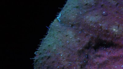


So what do we see here? A mix of blueish fluorescent emitted light, mainly from these little hairs on the surface of the leaf and from the stomata which allows the leaf cells to exchange gases (Carbon Dioxide and Oxygen) with the surrounding atmosphere.


The reddish NIR flourescence seem to come from deeper inside the cells, no wonder actually, since it is a reaction of the chlorophyll deep within the plant cells (contained in the cell chloroplasts)! 


Sigh - the ever present lint! Here now a shot from the middle of the leaf:


So I hope you enjoyed that high resolution journey to a Primula leaf!


Stay tuned, more will follow on that fascinating subject...

More info on this very interesting field may be found on my site http://www.pbase.com/kds315/uv_photos

Primula III ... Multispectral Imaging


So, that Primula again. How boring? Guess not ... since today it will be more than "just" that effect of stimulated IR flourescence, it will be about Multispectral Imaging also. And that means combining shots done in different wavelengths of light (not necessarily visible light) into just one image. So this is how that well known flower looks like in visible light:

[as usual, a click on an image opens up a larger view]


Under UV light, using a combination of Xenon and a 365nm Nichia UV LED flash/lamp, the scene now looks like this. Not much UV reflection actually, this is why it looks a bit dull. If you look more carefully, you might notice how UV light enhances even the smallest details and especially shows damages to the sensitives flower petals, like nicks and bruises (a reason why UV photography is used in forensics with great success, since even older marks which might not yet or no longer be visible, would still show under UV light).


Using the same UV light, but an UV stopping filter in front of the X35 lens, in this case the 2" Baader UV/IR Cut filter, the scene now changes since it is about UV stimulated flourescence what we see. The pollen lights up greenish/blueish and that deep red lights up from the green leaves actually is red IR flourescence (stimulated by UV).


Changing back to visible light, but using a deep red IR filter, yields the following result (not that spectacular, since there is not much IR response):


If we now map three of the images above into one resulting image via UV --> B, VIS --> G, IR --> R channels and combine these into one image, we get:


Which now shows three different wavebands combined in just one image.

So I hope you enjoyed that "multispectral" Primula flower!

Stay tuned, more will follow on that fascinating subject...

More info on this very interesting field may be found on my site http://www.pbase.com/kds315/uv_photos