Showing posts with label UV. Show all posts

Sunday, November 16, 2014

Enhancing the visibility of fossil tissue structures using UV reflected and UV stimulated visible flourescence photography

A while ago I had the honor to work with Neal Larson on a paper on fossilized cephalopods found in Hajoula, Lebanon. Today it is again about enhancing the visibility of fossil bone and tissue structures using UV reflected and UV stimulated visible fluorescence photography, but as it is some years later, using more modern equipment now. I will also use my remapping technology consisting of a visible image, a reflected UV image and an UV stimulated visible fluorescence image and combine them into multispectral images.

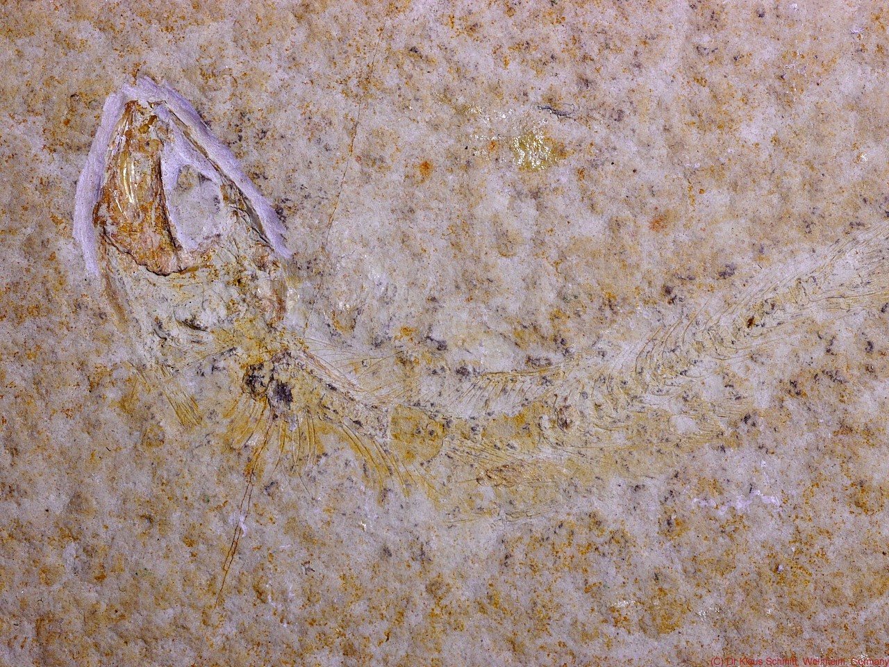
Lens used was my CERCO f4.1 / 94mm quartz fluorite lens, light sources were a modified high power Xenon flash, as well as a NICHIA 365nm Power LED. Target was a fossilized fish from Solnhofen, Germany, approx. 100 Mio years old.

[click on image to see a larger one]

Visible light image using UV/IR Cut filter:

Reflected UV image using Baader-U filter (310-390nm): 


UV stimulated visible fluorescence (FL) using Nichia 365nm UV LED:


Combined VIS - FL multispectral image: 


Combined VIS - UV multispectral image::


Combined FL - UV multispectral image::


It gets nicely visible that using UV light photography brings out much more details than normal visible light photography and by doing so, enhances the visibility of preserved bone and tissue structures quite a bit. Combining those different images into falso color multispectral images  enhances the structures even more.

Stay tuned, more will follow on that fascinating subject...

More info on this very interesting field may be found on my site http://www.pbase.com/kds315/uv_photos

Tuesday, September 16, 2014

Making of Thomas Leveritt video "How the sun sees you" - reflected ultraviolet (UV) photography part I

Well, in August 2014 a video "How the sun sees you" by Thomas Leveritt went viral on the net, a video about using sun cream for skin cancer protection and raising awareness about the need for skin protection. Leveritt used materials and techniques which were known by UV photographers since many years and my readers here have seen my older videos which employed the very same techniques, however used to make visible what bees, butterflies and other insects see and we cannot - ultraviolet (UV) light. Usually I use a false color palette to make UV visible.

Have a look at the following HD black+white video clip to see that it is indeed doable. I would like to show in part II later that such a video can be made using a suitable camera, lens + filter.


Now the following is the same HD video clip, but this time in false UV colors. They have a meaning, but this might be revealed later on.


I hope you enjoyed this funny little clip today!

Stay tuned, more will follow on that fascinating subject...

More info on this very interesting field may be found on my site http://www.pbase.com/kds315/uv_photos

Monday, April 9, 2012

[UV] Light Source - Flower - Filter - Lens - Sensor: a simulation of reflected UV photography

Well, just some ideas that came into my mind quite a while ago, when I was thinking about the chain:
Light Source -> Flower -> Filter -> Lens ->Sensor

To simplify things, let's assume

a) the light source being sunlight or Xenon light, as they are quite similar in the interesting UV region 300-400nm.
b) the lens being a quartz fluorite lens with flat transmittance in the interesting UV region 300-400nm

[click on image to see a larger one]

So here graphs (normalized to unity) showing in this example
1) flower (Mexican Zinnia, UV reflective flower tip): green line
2) filter (Baader U filter 2"): blue line
3) sensor (Nikon D200 mod. with quartz glass internal filter): red line


now lets add what that flower looks like after having passed the filter (pink line)


and finally what that sensor records of that (cyan line)


btw. if a normal, but UV transmitting glass lens (Noflexar 35mm - yellowish line) is used between filter and sensor, then the result looks like that:


The simple laymans terms summary: This is what the flowers reflects (green line) and what the camera records of it (cyan line) through a Baader U filter and Noflexar 35mm lens:


The bad news is, that it will only be a bit better if a quartz fluorite lens is being used, but not substantially.


So in the first case the 320-390nm Baader U filter appears like a 372BP15 bandpass filter and in the second, better example, like a 365BP16 bandpass filter. This has nothing to do with the type or quality of the Baader U filter, but is caused only by the process chain it is used within!


***THESE ARE VERY SIMPLIFIED SIMULATIONS BARE SCIENTIFIC ACCURRACY, SO NO DECISION, JUST INSIGHT SHOULD BE BASED ON THAT***


Stay tuned, more will follow on that fascinating subject...

More info on this very interesting field may be found on my site http://www.pbase.com/kds315/uv_photos

Wednesday, November 17, 2010

Aster hybride meets Noflexar 35mm lens

Today few shots of an Aster hybride using the Noflexar 3.5/35mm lens and various filters.
No noticeable patterns, as expected though.

[click on image to see a larger one]

visible light shot: 


UV shot (using Baader U-filter and Xenon flash): 


Butterfly vision shot (using my XBV2 filter): 


UV induced visible flourescence shot (using Baader UV/IR Cut filter and Nichia 365nm UV light): 


As boring as it might look like, this now is quite interesting (to me) as it shows UV induced IR fluorescence. Lights were all Nichia UV LED sources I could get hold of (to make sure not a bit of IR would be present), a B+W 092 cut filter (>65nm) in front of the lens and 30sec exposure in total darkness:


With the naked (but UV protected!!) eye quite a bit deep red fluorescence gets visible, but hard to ban that on a photo....

Same experiment but using a dark orange filter and same UV lighting setup in darkness (Nichia 4x 365nm LED using a front attached Baader U-filter to block any visible light!):


I have now found a reliable method to professionally infinity convert Noflexar 35mm lenses to Nikon mount - ask me if you need one!

Stay tuned, more will follow on that fascinating subject...

More info on this very interesting field may be found on my site http://www.pbase.com/kds315/uv_photos

Thursday, July 15, 2010

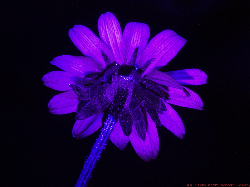
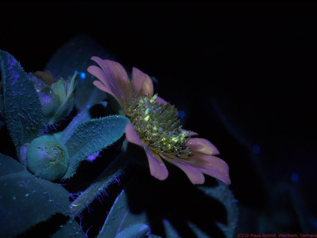
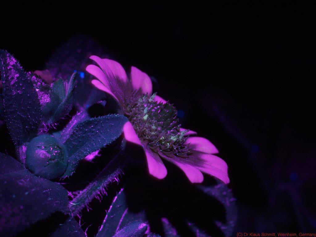
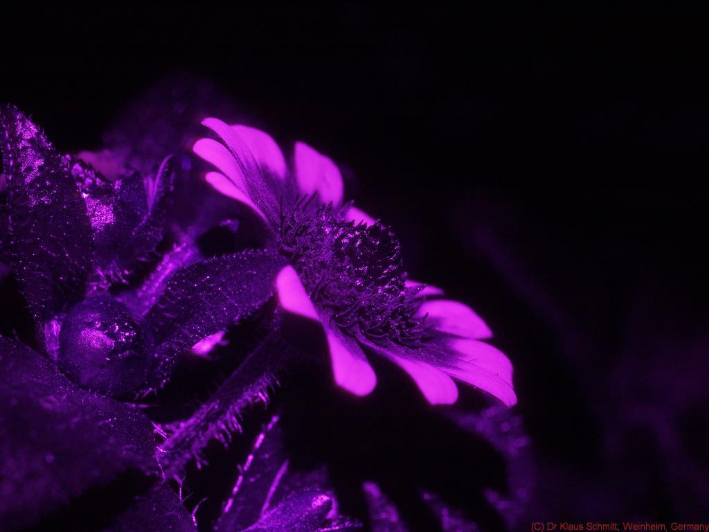
Rudbeckia hirta flower bottom up

This is again about a Rudbeckia hirta a flower but this time shot bottum up.

[click on image to see a large image]

UV LED lit showing visible NIR fluorescence 


butterfly vision image shot using Xenon light:


Visible image shot using Xenon light 


Visible stereo image (for crosseyed viewing)


Stay tuned, more will follow on that fascinating subject...

More info on this very interesting field may be found on my site http://www.pbase.com/kds315/uv_photos

Saturday, April 17, 2010

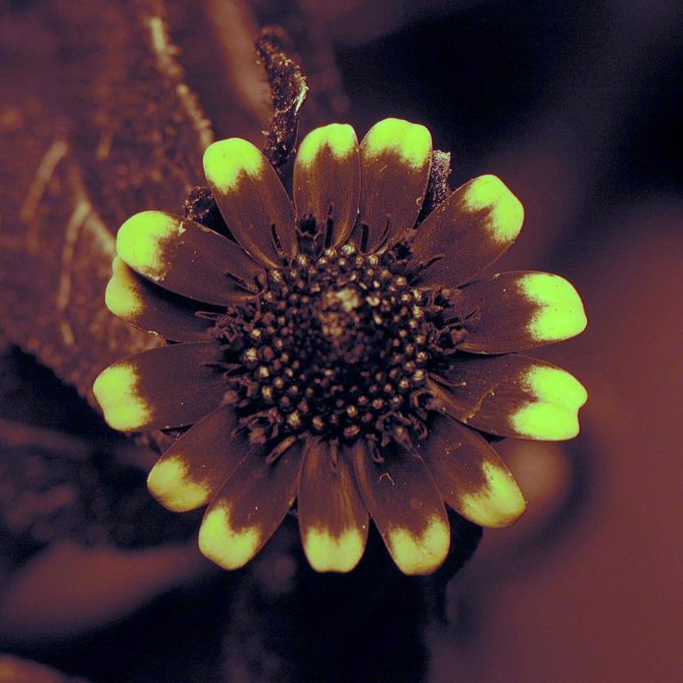
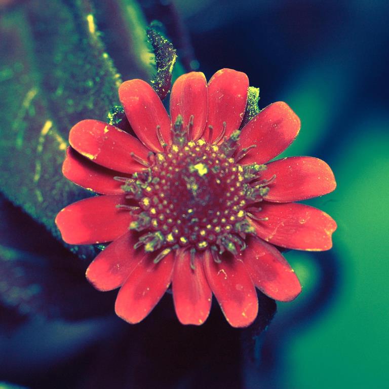
Creeping Zinnia multispectral

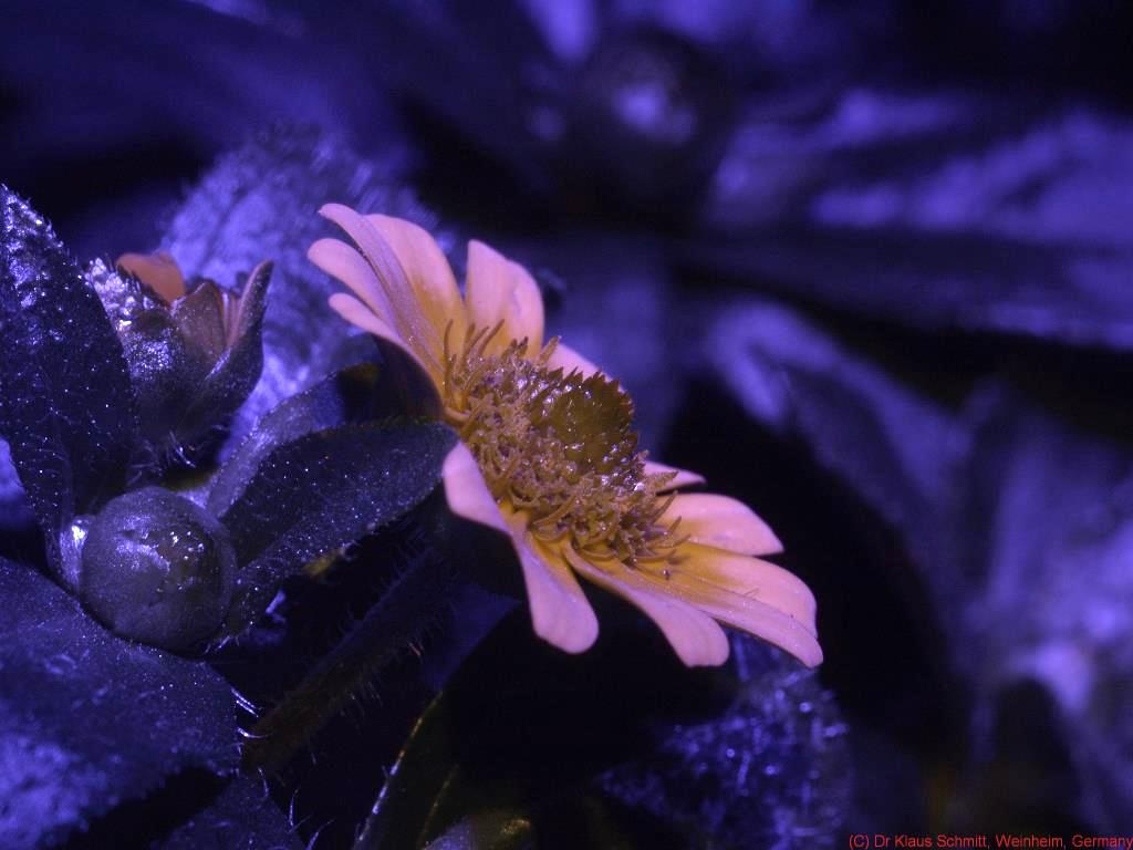
The first Creeping Zinnias appeared in 2010, so I took a chance to shoot that interesting little flower using a UV-Planar4/60mm lens and various filters and lightsources.

[click on image to see a larger one]

Visual shot 


FL (UV induced visilble fluorescence) shot 


UV+FL shot 


UV (300-400nm) shot 


BV (simulated bee vision) shot 


Stay tuned, more will follow on that fascinating subject...

More info on this very interesting field may be found on my site http://www.pbase.com/kds315/uv_photos

Sunday, April 11, 2010

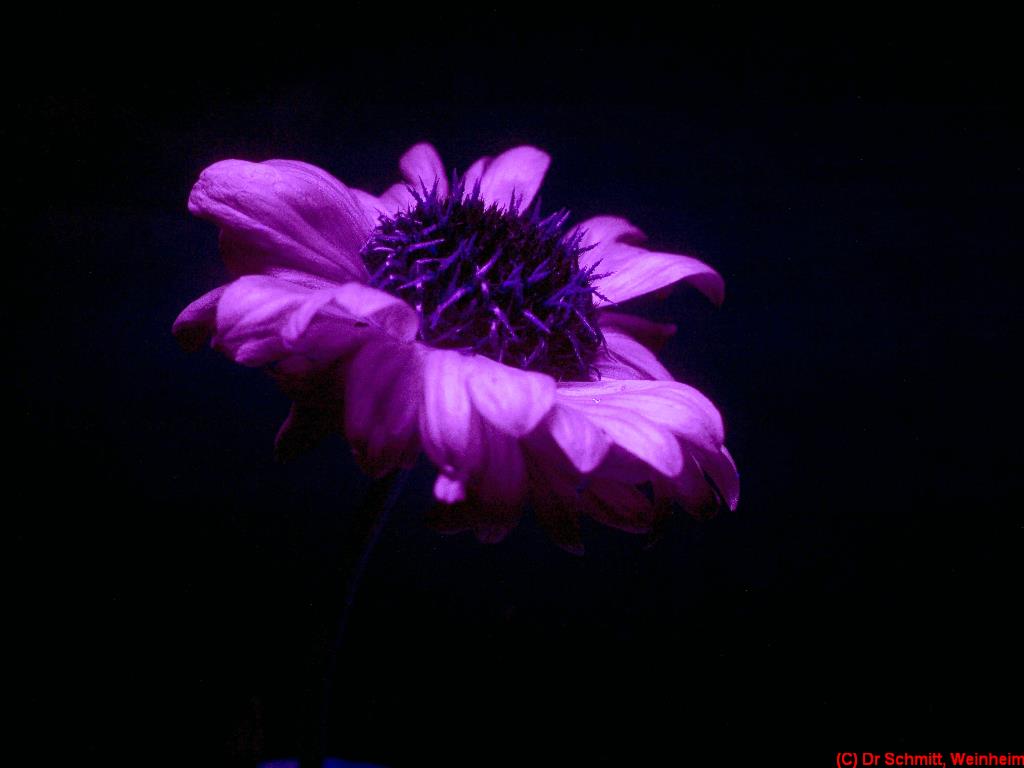
[FL] Another Bouquet: Gerber Daisy, Hyacinth, Roses,...

Here a UV induced visible fluorescence shot of another flower bouquet using Nichia 365nm UV LED and a 400-700nm UV/IR Cut filter.

[click on image to see a larger one]


and here for comparison the white light (5600K LED) shot


closer to see more details...


Well and here as another comparison, what happens when a Xenon flash with UV filter is used to stimulate fluorescence.

UV Xenon flash, but that leads to IR + red contamination as may be seen here:


and here now using a BG filter to compensate for that


but in overall comparison, the Nichia UV LED wins hands down IMHO!


Stay tuned, more will follow on that fascinating subject...

More info on this very interesting field may be found on my site http://www.pbase.com/kds315/uv_photos

Tuesday, March 30, 2010

[UV, FL] Enhancing the visibility of fossil cephalopods soft tissue structures

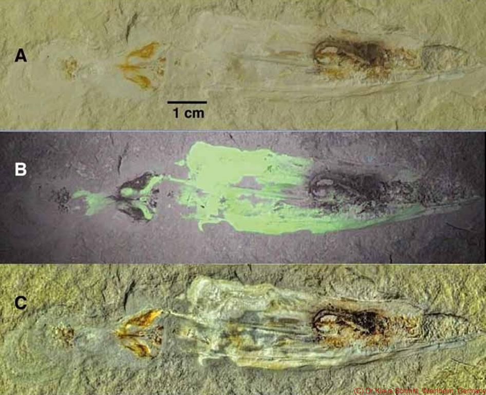
I had the honor to work with Neal Larson (yeah, THAT Neal Larson who discovered SUE, the T-Rex, with his brother Peter) on a paper on fossilized cephalopods found in Hajoula, Lebanon. I used my remapping technology consisting of a visible image (A), a UV stimulated visible fluorescence image (B) to enhance the visibility of preserved soft tissue structures (C) by combining them into one multispectral image.

Should someone be interested in the paper, the full title is:
"A new look at fossil cephalopods", NEAL L. LARSON, ROBERT W. MORTON, PETER L. LARSON AND UWE BERGMANN in
Tanabe, K., Shigeta, Y., Sasaki, T. & Hirano, H. (eds.) 2010. Cephalopods - Present and Past, Tokai University Press, Tokyo, p. 303-314.

Images (c) Neal L Larson and Dr Klaus Schmitt

[click on image to see a larger one]


Another example of the same technique, shots I did with a squid fossil I have here from the Upper Cretaceous period, ca 90 million years old.

Visible light image:

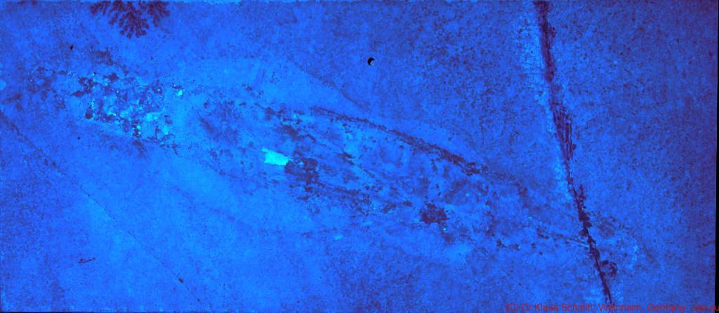
UV stimulated visible fluorescence: 

multispectral remapped 


There is a follow up to this one HERE.

Stay tuned, more will follow on that fascinating subject...

More info on this very interesting field may be found on my site http://www.pbase.com/kds315/uv_photos
Friday, July 10, 2009

Gaillardia "Summerwind"

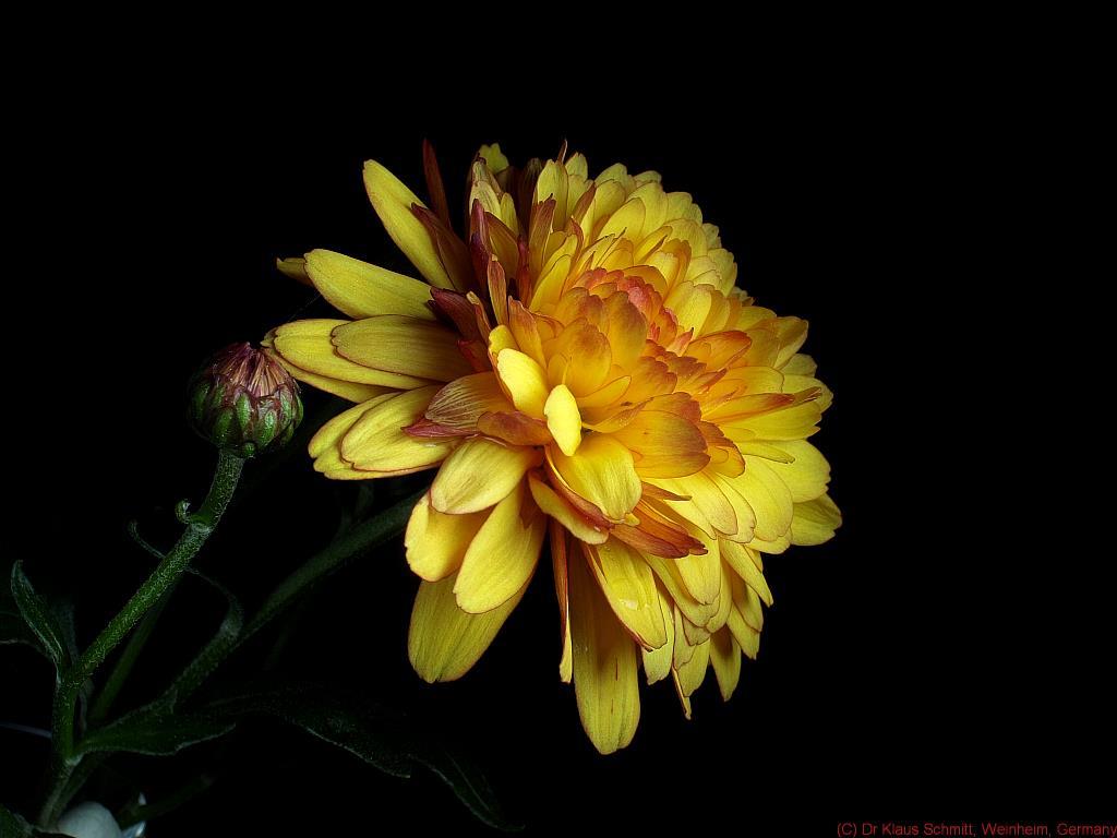
Well, I found a previously unknown (to me), pure yellow species of Gaillardia. Since that type of flower showed so many surprising aspects, I wanted to see what this new one hides from the human eye....

[click on image yields a larger one]

This now is the visual appearance as we humans are used to see:


The tetrachromatic image ("butterfly" shot) (UV+B+G+R) using XBV2 filter:


The trichromatic image ("bee vision" shot) (UV+B+G) using XBV2 filter:


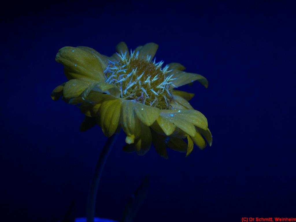
This is the UV induced fluorescence shot using XCUT2 filter:


and this the reflected UV shot using XUVIR filter:


Well, also that flower shows some surprising properties ...

Stay tuned, more will follow on that fascinating subject...

More info on this very interesting field may be found on my site http://www.pbase.com/kds315/uv_photos

Monday, April 20, 2009

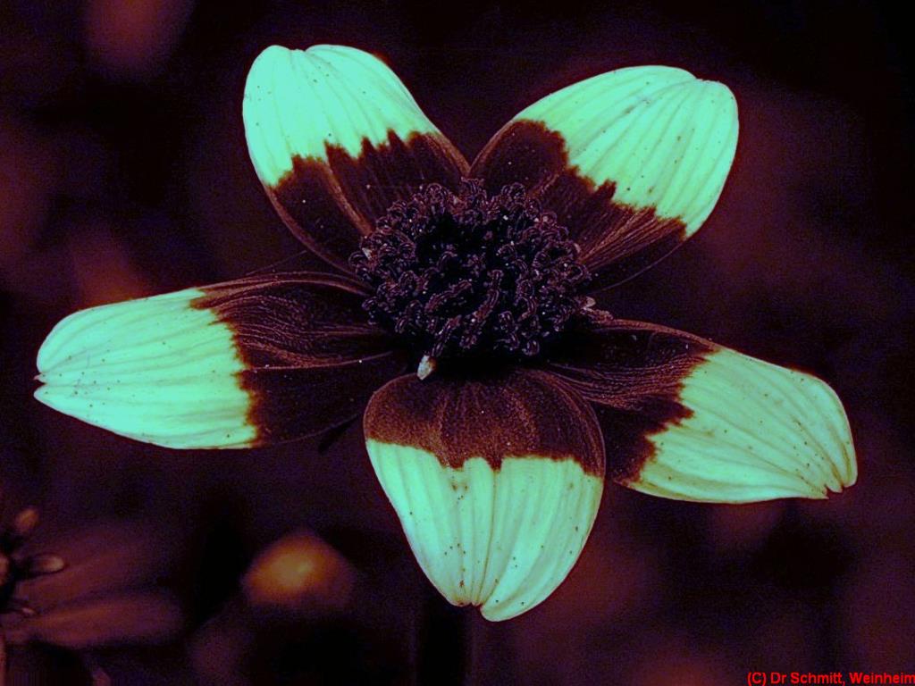
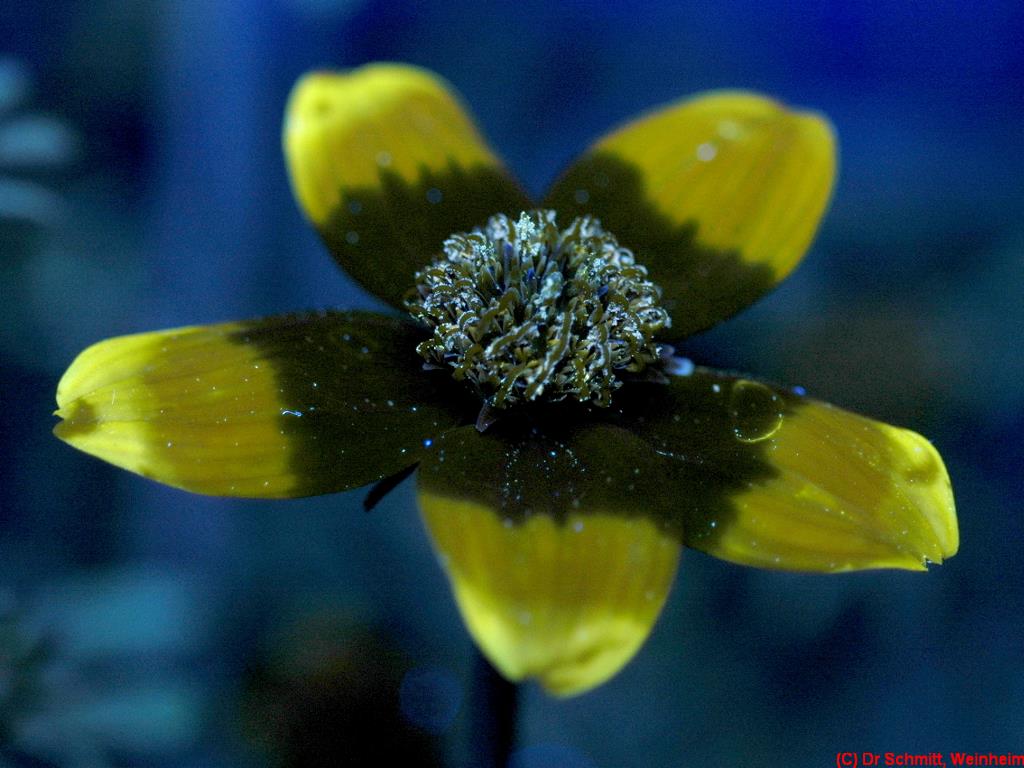
Bidens ferulifolia, various filters + light sources

Well, another nice target for UV photographic experiments is Bidens ferulifolia, the variety "Peters's Surprise".

Equipment used was: Nikon D70 (unmodified), ISO400, f5.6-8, 1/160sec, old 100mm enlarger lens.

[click on image to see a larger one]

VIS using XCUT2 filter (400-650nm):


UV using Baader U-filter (310-390nm) - synthetic DOF:


XUVIR multispectral filter (300-1000nm), UV Flash:


UV induced fluorescence using XCUT2 filter (400-650nm), UV flash:

So, as you may see, a variety of results also that flower shows us.

P.S.: if you wonder what "synthetic DOF" means, I used "stacking" to increase the DOF artificially.


Stay tuned, more will follow on that fascinating subject...

More info on this very interesting field may be found on my site http://www.pbase.com/kds315/uv_photos
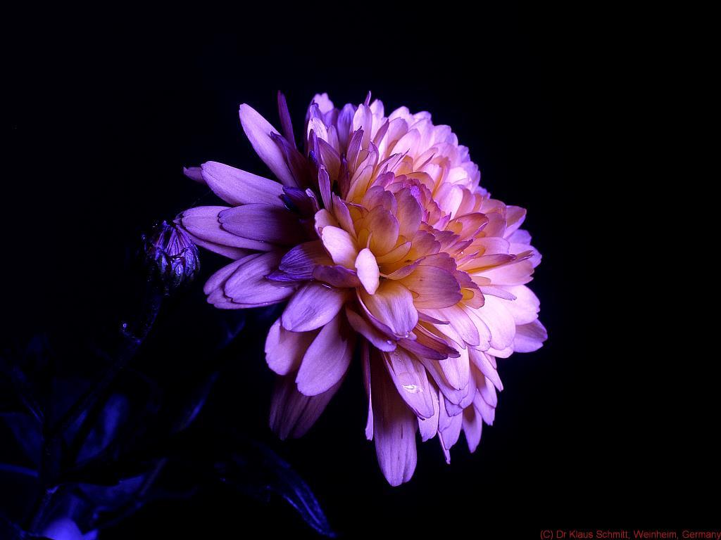
Thursday, April 16, 2009

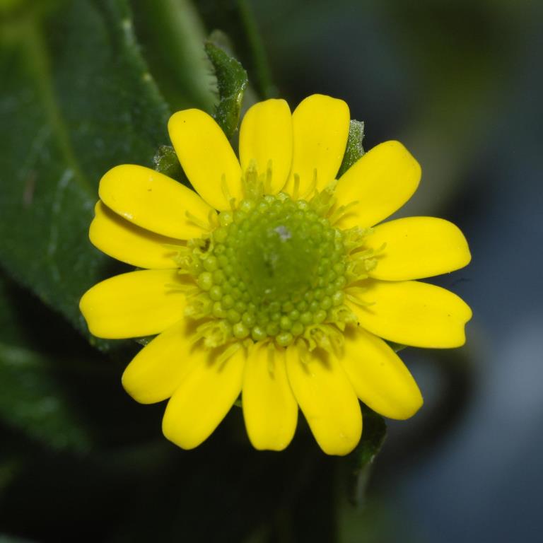
Creeping Zinna, multispectral, UV induced fluorescence, reflected UV etc.

A walk over the local market not only brought some nice white "Lambertheimer" asperagus and new "Galatiner" potatoes home, nope, also a nice "Creeping Zinna" (Sanvitalia procumbens) plant made it to my balcony.

An ideal target to test out the UV enhanced High Power Xenon, the XUVIR "one shot" filter, the Xcut2 filter as well as the well known Baader U filter.

Equipment used was: Nikon D70 (unmodified), UV Nikkor 105mm lens @ ISO400, f11, 1/180sec.

[click on image to see a larger one]

VIS using XCUT2 filter (400-650nm):


UV using Baader U-filter (310-390nm):


XUVIR multispectral filter (300-1000nm), UV Flash:


XUVIR filter (300-1000nm), unfiltered flash for multispectral recording


UV induced fluorescence using XCUT2 filter (400-650nm), UV flash:


Sanvitalias UV pattern is actually not that easy to shoot. That may been seen if you have a look at the reflection spectra which I took, since the UV reflection of the petal tip (white line) is less than 10%, just a little bit more than the rest of the petal (teal line) which has hardly any UV reflection. At the same time the IR reflection is very high. So very careful filtering and a strong UV source is needed :


So, as you may see, a variety of results may be produced....and yes, the asparagus was delicious...

Stay tuned, more will follow on that fascinating subject...

More info on this very interesting field may be found on my site http://www.pbase.com/kds315/uv_photos