While I was shooting flowers at Herrmanshof, Weinheim park, I noticed some intense whiteish and some dimmer greenish UV reflectance patters (in my standardized false UV color palette) and I wanted to have some proof, if from the false color UV displayed it could be backward deducted about a specific
sub-UV-A waveband reflection using spectrometric measurements .
[click on images to see larger ones]
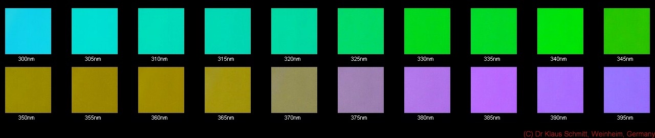
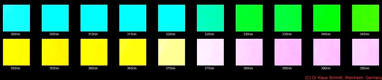
Standardized reflected false UV "colors" according to the previously described "
UV color palette" related to wavelength.
The normal intensity palette:

and this is the high intensity palette:

Well, may I remind, that these false colors do not co-exist at the same time with the same intensity at the same wavelength stimulus (hence cannot be mixed), because of the very different sensor sensitivity to different UV wavelengths.

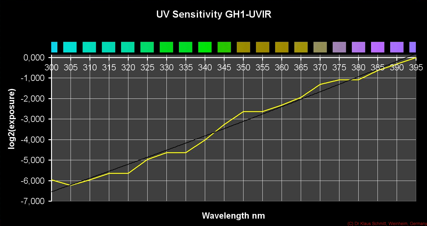
So from that above re-posted graph, which shows that GH1-UVIR sensor reaction to a monochromatic 5nm bandwith stimulus, for instance a 340nm "UV color" needs 4EV more exposure than one at 395nm and a 300nm needs even 6EV more.
This is why I found it so special to find a green response (340nm) and yellowish response (370nm) in the same image (Zinnias as shown; also the reason why the green is so dim and the whitish yellow so overexposed) as there is 3EV in between - and this is why I doubt to ever find sea-green (around 300nm) in such a photo (6EV difference) that would contain this violet (at 395nm) - except I use a special technique which already forms in my mind...

So if one wanted to simulate how the UV color would look like in a resulting photo, we could use the spectrometer output and would have to multiply it with the Baader-U filter transmittance actually and then look up the exposure values per intensity. Unfortunately the sensor also responds nonlinear over wide exposure ranges (so the above is valid for mid level exposures only), so that yields only a crude approximation. But at least one would get a clue why that petal tip appeared green - yellowish in that image as posted above.
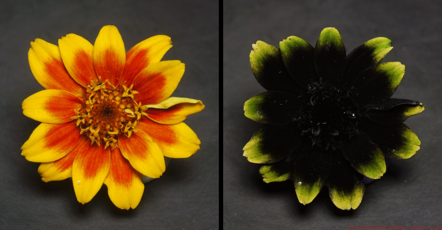
Spectrometric result of such a petal of the following Zinnia variant, tip greenish, rest nearly black (in UV) and yellow tip, orange-red the petal rest in VIS:

Photographic result: VIS:left, UV:right

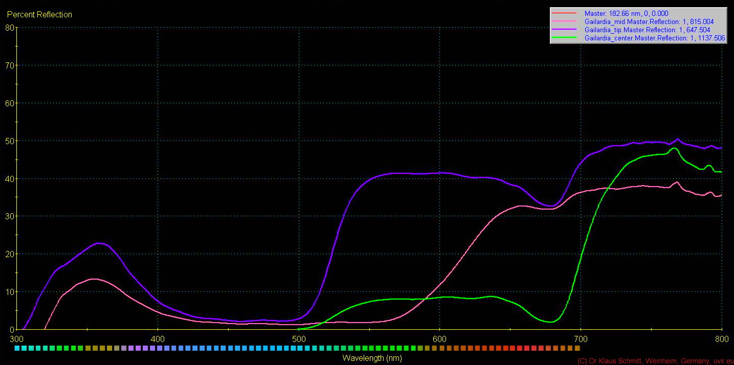
Following examples for comparison,: Bidens ferulifolia, petal tip looks bright yellowish and rest petal quite dark in UV and Sanvitalia procumbens which looks about the same UV color at its petal tip but dimmer and petal rest nearly pitch black in UV (using my standardized false UV color palette). Gaillardia aristata has an underlying UV reflective layer even over the whole tip under a in the visible yellow petal tip and orange-red rest of the tip; the flower center is all UV dark but in the visible orange red outside and greenish in the very center
Spectrometric results
Bidens ferulifolia:

Prediction "UV color": bright whiteish-yellowish tip, dark rest of petal; dark center
Sanvitalia procumbens:

Prediction "UV color": whiteish-yellowish tip, dark rest of petal; dark center
Photographic result: UV (right part of image is sunlight, left 365nm UV LED; Bidens left, Sanvitalia right):

Gaillardia aristata:

Prediction "UV color": bright whiteish-yellowish petal; dark center
Photographic result: VIS / UV (wet, after rain):

Added today:
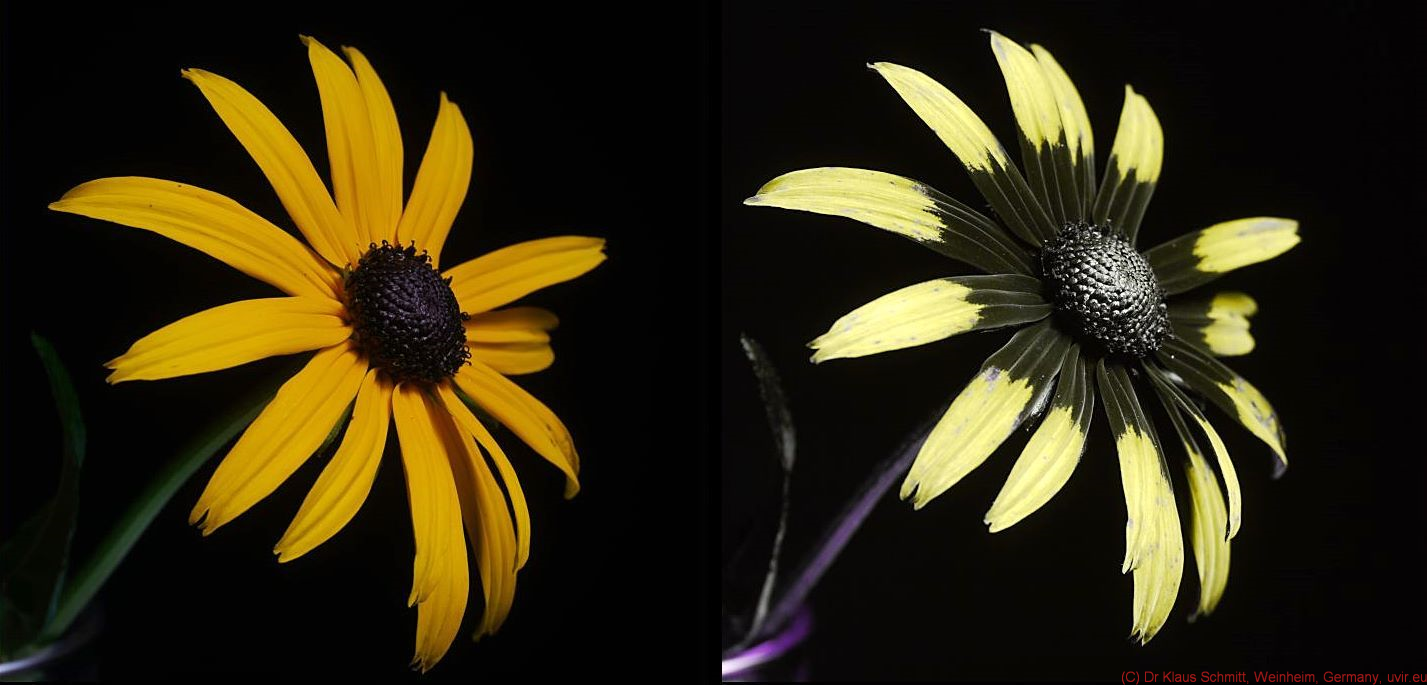
Rudbeckia fulgida flower:

Prediction "UV color": yellow tip, dark rest of petal; whiteish-yellowish center reflection
Photographic VIS/UV:

So we see, that it holds true so far, that my standardized false UV colors can stand for specific UV-A sub-waveband reflections as shown
here on my BLOG.
Stay tuned, more will follow on that fascinating subject...
More info on this very interesting field may be found on my site
http://www.pbase.com/kds315/uv_photos