Wednesday, April 9, 2008

Bumble Bee on Mexican Zinnia

Today just one image I wanted to share, a Bumble Bee on a Mexican Zinnia (?) flower - how boring? Not really, since both exhibit their UV patterns; have a look....

Also these two shots were done using extention tubes and a Baader 2" U-filter for the UV shot using my Nichia 365nm UV Led lamp/flash. Now here they come assembled into one image and the UV image somewhat matched in color to enhance the UV pattern:


Notice the detail the UV shot has in comparison to the white light image. Quite easy to understand actually, since the (theoretical) resolution of that lens used rises by a factor of 546nm/365nm = ca 1.5x (if we use the median for white light).

So since I had a feeling someone might ask how a bee might see that situation, here my sythesized bee vision version for your viewing pleasure:


Stay tuned, more will follow on that fascinating subject...

More info on this very interesting field may be found on my site http://www.pbase.com/kds315/uv_photos

Monday, March 24, 2008

Easter Daffodil

It is Easter and the Daffodils are in full bloom. I wondered how might these look like under ultraviolet light?

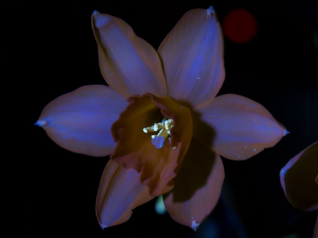
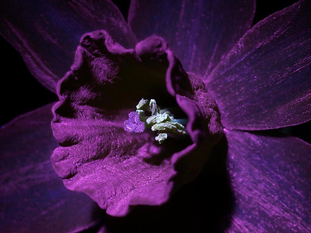
These shots were done using an extention tube, an UV Rodagon 60mm and a Baader UV/IR Cut filter to suppress the UV exciting light source (Nichia 365nm UV Led lamp/flash). Here the visible shot first. [as usual, a click on an image opens up a larger view]


And now how this flower looks like when radiated with UV light @365nm. The pollen shines bright in UV induced fluorescent yellowish white, whereas the petals emit some reddish and blueish colors. Especially the tips shine up very bright.


Now on to the reflected UV light image here. Not much reflectance to notice though, but a beautiful velvetish surface!


So I hope you enjoyed that journey to an Easter Daffodil with me!


Stay tuned, more will follow on that fascinating subject...

More info on this very interesting field may be found on my site http://www.pbase.com/kds315/uv_photos

Sunday, March 9, 2008

Primula IV - a high Resolution Journey

More on Primula and its secrets - this time much we have a much closer look at that mysterious red UV induced NIT fluorescence.

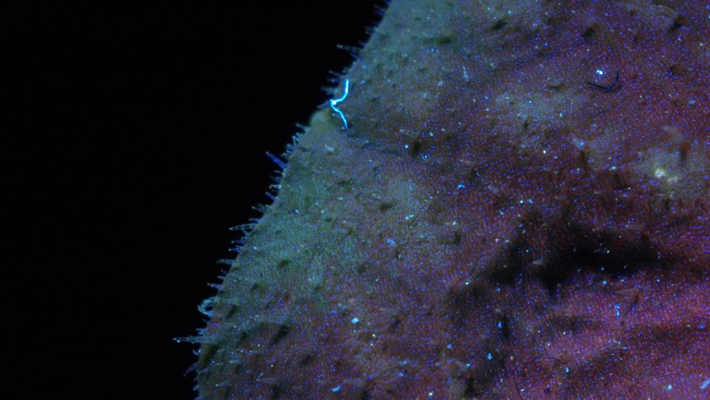
These shots were done using a long extention tube, a UV Rodagon 60mm and a Baader UV/IR Cut filter to suppress the UV exciting light source (Nichia 365nm UV Led lamp/flash). Let's have a look at a green leaf here: [as usual, a click on an image opens up a larger view]


So what do we see here? A mix of blueish fluorescent emitted light, mainly from these little hairs on the surface of the leaf and from the stomata which allows the leaf cells to exchange gases (Carbon Dioxide and Oxygen) with the surrounding atmosphere.


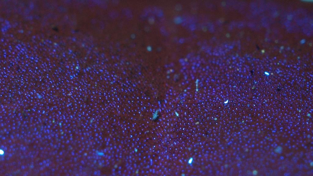
The reddish NIR flourescence seem to come from deeper inside the cells, no wonder actually, since it is a reaction of the chlorophyll deep within the plant cells (contained in the cell chloroplasts)! 


Sigh - the ever present lint! Here now a shot from the middle of the leaf:


So I hope you enjoyed that high resolution journey to a Primula leaf!


Stay tuned, more will follow on that fascinating subject...

More info on this very interesting field may be found on my site http://www.pbase.com/kds315/uv_photos

Primula III ... Multispectral Imaging


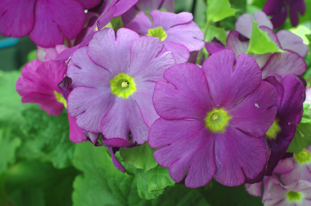
So, that Primula again. How boring? Guess not ... since today it will be more than "just" that effect of stimulated IR flourescence, it will be about Multispectral Imaging also. And that means combining shots done in different wavelengths of light (not necessarily visible light) into just one image. So this is how that well known flower looks like in visible light:

[as usual, a click on an image opens up a larger view]


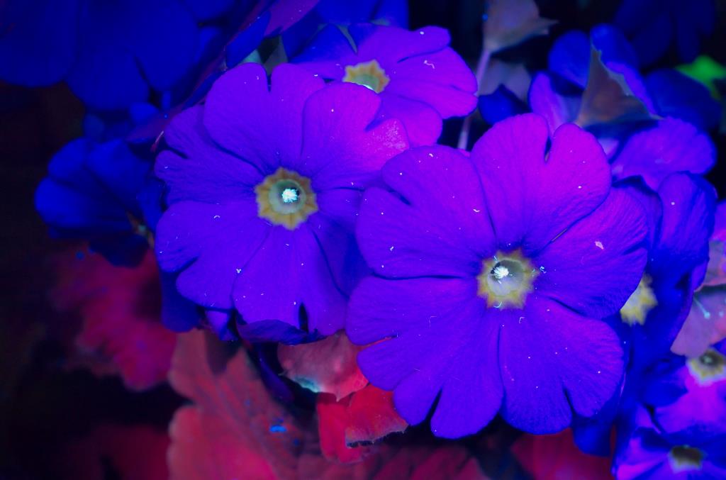
Under UV light, using a combination of Xenon and a 365nm Nichia UV LED flash/lamp, the scene now looks like this. Not much UV reflection actually, this is why it looks a bit dull. If you look more carefully, you might notice how UV light enhances even the smallest details and especially shows damages to the sensitives flower petals, like nicks and bruises (a reason why UV photography is used in forensics with great success, since even older marks which might not yet or no longer be visible, would still show under UV light).


Using the same UV light, but an UV stopping filter in front of the X35 lens, in this case the 2" Baader UV/IR Cut filter, the scene now changes since it is about UV stimulated flourescence what we see. The pollen lights up greenish/blueish and that deep red lights up from the green leaves actually is red IR flourescence (stimulated by UV).


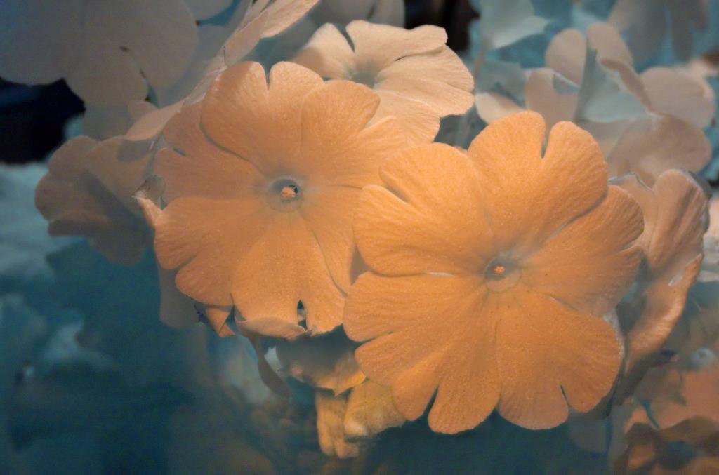
Changing back to visible light, but using a deep red IR filter, yields the following result (not that spectacular, since there is not much IR response):


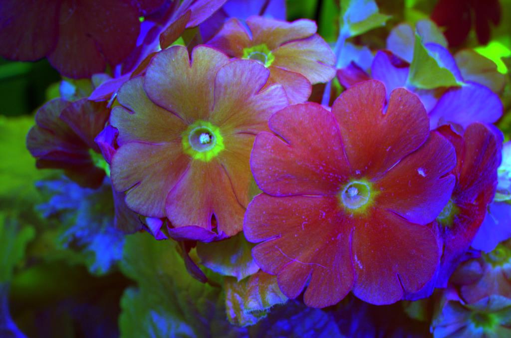
If we now map three of the images above into one resulting image via UV --> B, VIS --> G, IR --> R channels and combine these into one image, we get:


Which now shows three different wavebands combined in just one image.

So I hope you enjoyed that "multispectral" Primula flower!

Stay tuned, more will follow on that fascinating subject...

More info on this very interesting field may be found on my site http://www.pbase.com/kds315/uv_photos

Sunday, March 2, 2008

Primula I ... NIR flourescence


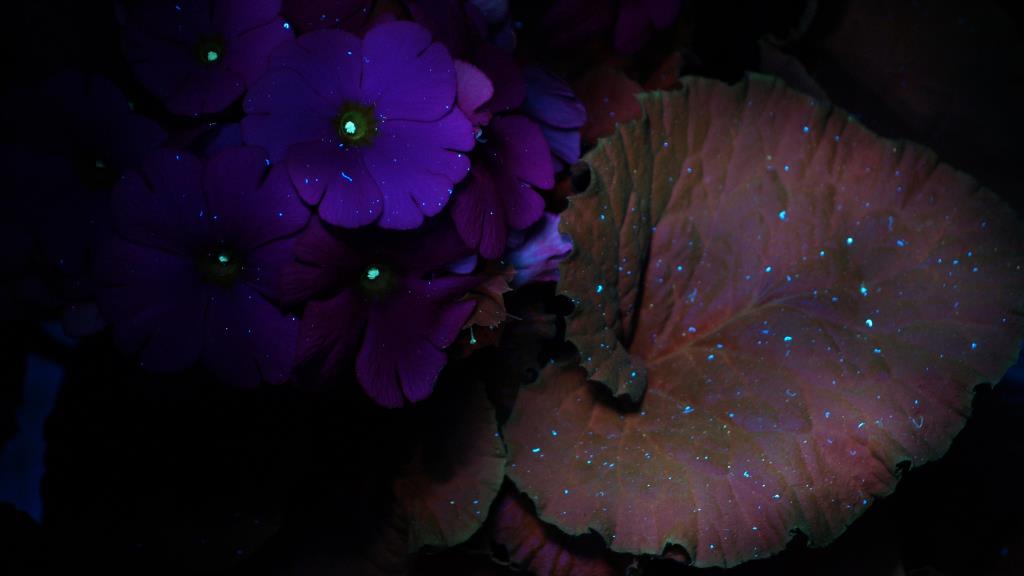
I wondered around in my home with my mobile UV LED Lamp in my hands, my UV stopping googles on in search of some unusual effects this strong 365nm UV light might show. A primula plant I just bought catched my attention, since while I was lighting the flowers (the "usual suspects" so to speak...], UV light also fell onto the deep green leaves of that beautiful plant. And to my big surprise, I saw them lighten up in a mysteriously dark glowing red!

I grabbed my camera and took some shots of that effect, using a suitable macro lens (X35) and a UV Cut filter - more about that later, since it turned out to be a nontrivial issue....

So this is how that plant looked in visible light, well known to most of us: [as usual, a click on an image opens up a larger view]


Switching from visible light to UV light, using my 365nm Nichia UV LED flash/lamp, the scene dramatically changes and that deep red lights up from the green leaves! I used an UV stopping filter in front of the lens, in this case the 2" Baader UV/IR Cut filter.


To record that emitted red light in a picture turned out to be nontrivial, since most cut filters I have here also cut off that red light and I just got a grey or greenish color. The spectrometer reveals why this is the case, it starts at about 650nm and extends into the near IR (NIR) region, most of these filter cut off. The only one which worked somewhat was the Baader UV/IR Cut filter. The Canon, Tiffen etc. UV/IR cut filters ("hot mirror") all failed since they are designed to cut off NIR.


But what is that what we see here? It is UV stimulated near infrared (NIR) fluorescence of the green chlorophyll (which is a type of porphyrine we have seen in the egg study I published earlier)!

So I hope you enjoyed that "hidden beauty" of that Primula flower!


Stay tuned, more will follow on that fascinating subject...

More info on this very interesting field may be found on my site http://www.pbase.com/kds315/uv_photos

Primula II ... UV induced visible Fluorescence

So today a flower fell of that beautiful Primula plant. I had bought that for my balcony, since I needed some colors around me after a long a dark winter, which is finally over here in Weinheim / Germany

I grabbed my camera, attached a long extention tube, plus that tack sharp UV Rodagon 60mm lens. This following shot was done using a tungsten cold fiber light: [as usual, a click on an image opens up a larger view]


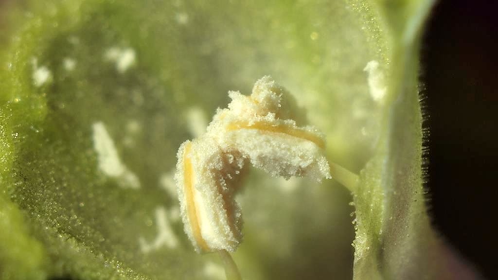
So if we switch from visible light to UV light, in this case using my 365nm Nichia UV LED flash/lamp, we will see the pollen light up blueish / greenish and also the colors of the flower petals change. I had to remember to use a UV stopping filter in front of the lens, in this case the 2" Baader UV/IR Cut filter, which also stops IR light very efficiently, needed for modern DSLRs due to their very high IR sensitivity. This is the result now, called UV induced visible fluorescence, commonly called fluorescence [there are other forms of fluorescence, too, like UV fluorecence, IR fluorescence just FYI].


So lets look a bit more in detail to these wonderful bright blueish/greenish lighting up pollen, first in visible light...


...then using UV light which stimulates flourescence. These shots need to be done in darkness, since that effect is quite weak and needs long exposure times:


So I hope you enjoyed having a look at a flower using UV induced visible fluorescence!


Stay tuned, more will follow on that fascinating subject...

More info on this very interesting field may be found on my site http://www.pbase.com/kds315/uv_photos

Friday, February 29, 2008

Easter is approaching...!

Easter is approaching and quite some shops exhibit the traditional easter decoration. I was curious to see, how that white ostrich egg would look multispectral.

[as usual, a click on an image opens up a larger view]


Now that made me curious and the idea was to investigate the behaviour in terms of fluorescence these eggs (their eggshell more precisely) might show.

A Baader 2" UV/IR Cut Filter was used for the visible and fluorescence light shots, a Baader 2" U-filter for the UV shots together with that calibrated X35 lens I made, the UV Nikkor 105mm and an unmodified Nikon D70.

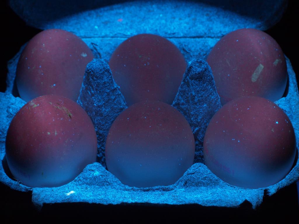
Now the big surprise is, that brown eggs exhibit some very unusual behaviour - they glow mysteriously red if you point a UVA light source to them; in that case here I used my mobile UV LED flash/lamp.


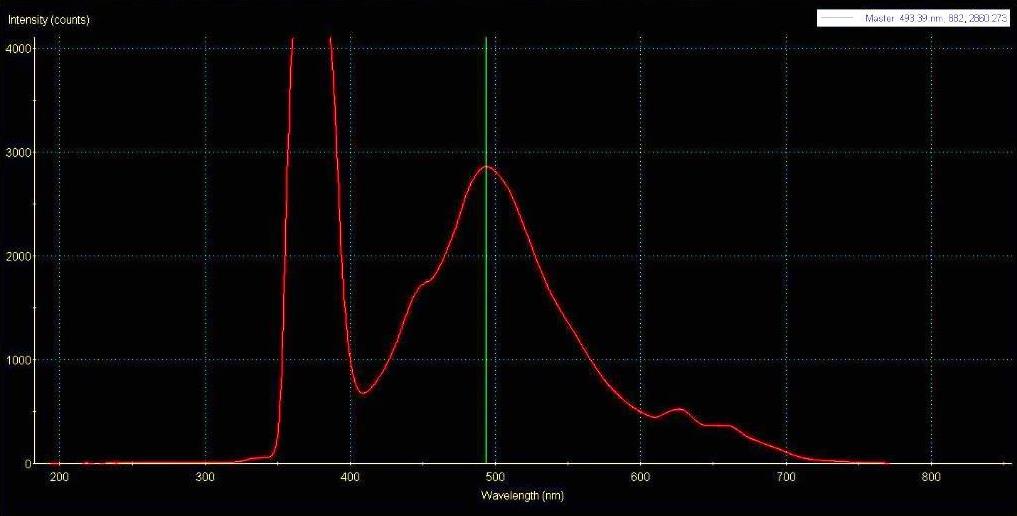
So why is that you might ask. It is UV stimulated visible red fluorescence of the porphyrine contained in the brown eggshells! This can be proven using a spectrophotometer (USB2000 in my case) which measures the reflected and emitted light from that eggshell with a fiber optic probe. The excitation source is quite strong, this is why that 365nm line is way off that graph. You see some response around 480nm (blueish) plus these two red porphyrine peaks at 675nm and a bit weaker 635nm.


Remember the first white ostrich egg shot I showed you above? I wondered if that one would also exhibit that effect, but the visual experience and the spectrophotometer reveals only a strong blueish response around 480nm plus a quite weak peak at around 635nm.


That made me even more curious and I got a fresh set of eggs from another vendor and indeed there it was again, that red fluorescence. Quite weaker but it was there, aside of some strong blueish fluorescence (as with that white ostrich egg):


The spectrometer confirmed these visual findings, the porphyrine response is clearly visible:


So I hope you enjoyed today this more scientific journey with me!


Stay tuned, more will follow on these fascinating subjects...

More info on this very interesting field may be found on my site http://www.pbase.com/kds315/uv_photos